Back Gone Out but Won’t Back Down
Author: Cassie Moylan, MD
Peer-Reviewer: Matthew Negaard, MD, CAQ-S
Final Editor: Alex Tomesch, MD, CAQ-SM
A 16-year-old male basketball, football, and soccer player presents with persistence of low back pain that began acutely 5-6 weeks ago during weight-lifting when he felt a sharp pain in his right lower back mid-descent in a squat.

Figure 1. Plain-films of the lumbar spine; AP (left) and Flexion (center) and Extension (right) Laterals. Author's own media, courtesy of Dr. Andrew Peterson, MD, MSPH.
What is the ED diagnosis? The most likely diagnosis?
The ED diagnosis is “low back pain”. The most likely diagnosis is spondylolysis. The patient’s plain-films are normal and in this condition they often will be. Additional imaging, obtained in follow up, is required for definitive diagnosis but not always necessary.
-
Pearl: Red flags necessitating emergent imaging in the ED include but are not limited to loss of sensation in the lower extremities, groin, and/or anogenital region; inability to stand; and inability to control bladder or bowel function.
Who is typically affected?
Spondylolysis is a fracture of the pars interarticularis that is most prevalent in adolescent athletes, up to 50% of those presenting with low back pain [1-5]. The L5 is the most commonly fractured of the vertebrae [1-3]. This condition may affect those as young as 6 years old but is most likely to occur during the adolescent growth spurt (between 10-15 years old) [4].
-
Pearl: Spondylolysis should always be on your differential for activity-limiting low back pain in an adolescent athlete, especially those participating in gymnastics, weight lifting, wrestling, and football (repetitive lumbar extension sports) [3].
-
Pearl: Pars interarticularis injury exists on a spectrum from stress reaction to fracture (spondylolysis) to vertebral slip (spondylolisthesis). Recognition of the diagnosis with proper management can prevent progression [1,3].
What physical exam findings are expected?
Patients may exhibit pain on palpation at the affected spinal level and with performing back extension. The Stork Test (single-legged hyperextension test) is often used to assess this diagnosis [1,2,4]. Patient’s do not typically have gross spinal deformity or neurologic findings on exam. They may have a variety of associated signs/symptoms including but not limited to paraspinal tenderness due to guarding, pain referred to the lower extremities, tight hamstrings and/or hip flexors, weak hip abductors and core musculature, and a posture of lumbar lordosis at baseline [1-4].
Which imaging modalities can be used?
In the absence of concerning signs or symptoms on exam, start with weight-bearing plain-films of the lumbar spine. You only need to obtain two views: standing AP and lateral. Lateral oblique views, described as best for viewing the pars interarticularis, are not necessary to obtain as they increase radiation without increasing sensitivity [1,2].
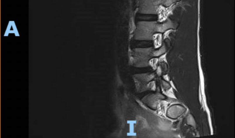
If plain-films indicate vertebral slippage or dynamic instability suggestive of spondylolisthesis additional imaging from the ED may be considered but is not always necessary. There is still debate about which imaging is the gold standard, but MRI is becoming very prevalent. CT and SPECT (previously considered gold standard) are still often used in certain cases. MRI is preferable given its ability to detect lesions with high sensitivity (under appropriate imaging protocols) and without radiation exposure in comparison to CT [1,2,4].

Figure 2. MRI of the lumbar spine demonstrating right pars interarticularis fracture of L5. Author’s own media, courtesy of Dr. Andrew Peterson, MD, MSPH.
Do you need to consult Ortho from the ED? What is the patient’s disposition from the ED?
No, orthopedics does not need to be consulted from the ED unless “red flags” of back pain are present. Referral to sports medicine or PCP is typically appropriate as these rarely require surgical intervention. Patient’s should be instructed to rest from sport and other pain-provocative activities until follow up.
References
[1] Chung CC, Shimer AL. Lumbosacral Spondylolysis and Spondylolisthesis. Clin Sports Med. 2021;40(3):471-490. doi:10.1016/j.csm.2021.03.004
[2] Stanitski CL. Spondylolysis and Spondylolisthesis in Athletes. Operative Techniques in Sports Medicine 2006;14(3):141-46 doi:https://doi.org/10.1053/j.otsm.2006.04.008[published Online First: Epub Date]|.
[3] Lawrence KJ, Elser T, Stromberg R. Lumbar spondylolysis in the adolescent athlete. Phys Ther Sport. 2016;20:56-60. doi:10.1016/j.ptsp.2016.04.003
[4] Berger RG, Doyle SM. Spondylolysis 2019 update. Current Opinion in Pediatrics 2019;31(1):61-68 doi: 10.1097/mop.0000000000000706[published Online First: Epub Date]|.
[5] Gregg CD, Dean S, Schneiders AG. Variables associated with active spondylolysis. Phys Ther Sport. 2009;10(4):121-124. doi:10.1016/j.ptsp.2009.08.001
[6] Alqarni AM, Schneiders AG, Cook CE, Hendrick PA. Clinical tests to diagnose lumbar spondylolysis and spondylolisthesis: A systematic review. Phys Ther Sport. 2015;16(3):268-275. doi:10.1016/j.ptsp.2014.12.005
[7] Masci L, Pike J, Malara F, Phillips B, Bennell K, Brukner P. Use of the one-legged hyperextension test and magnetic resonance imaging in the diagnosis of active spondylolysis. Br J Sports Med. 2006;40(11):940-946. doi:10.1136/bjsm.2006.030023
[8] Mellado JM, Larrosa R, Martín J, Yanguas N, Solanas S, Cozcolluela MR. MDCT of variations and anomalies of the neural arch and its processes: part 1--pedicles, pars interarticularis, laminae, and spinous process. AJR Am J Roentgenol. 2011;197(1):W104-W113. doi:10.2214/AJR.10.5803
[9] Tatsumura M, Gamada H, Okuwaki S, et al. Union evaluation of lumbar spondylolysis using MRI and CT in adolescents treated conservatively. J Orthop Sci. 2022;27(2):317-322. doi:10.1016/j.jos.2021.01.002


