Pelvic Injury
Author: William Perkins, MD
Peer-reviewer: Victor Huang, MD, CAQ-SM
Final Editor: Alex Tomesch, MD
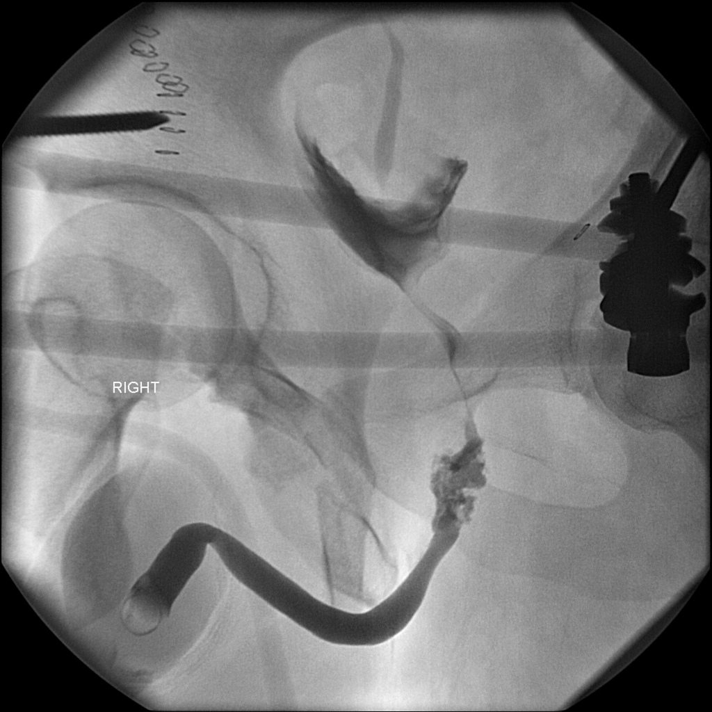
A 45 year old man presents to the Emergency Department with severe suprapubic pain after being thrown off of a forklift at work. On examination, there is significant pelvic tenderness to palpation, and blood at the urethral meatus.


Image Prompts. Image 1: AP view of the pelvis. Case courtesy of Dr. Andrew Dixon, Radiopaedia.org, rID: 31674. Image 2: Retrograde urethrogram. Case courtesy of Dr. Andrew Dixon, Radiopaedia.org, rID: 31648.
What is the diagnosis?
Bilateral pubic rami fracture with urethral injury seen on the retrograde urethrogram.
-
Pearl: Pelvic fractures have a mortality of 10-13%, and up to 50% if unstable [1]. Pelvic ring fractures are often associated with venous plexus or arterial injury [2].
-
Pearl: The Young-Burgess classification system differentiates pelvic ring fractures into three patterns based on the primary mechanism as well as the extent of injury and instability: Lateral Compression (LC), Anterior-Posterior Compression (APC), and Vertical Shear (VS) [1].
What is the mechanism of injury?
Pelvic fractures are seen in a bimodal distribution, with young men more likely to suffer from high energy trauma such as motor vehicle collisions and elderly females predisposed to osteoporosis more likely to suffer pelvic fractures after a low energy mechanism such as fall from standing [3].
What physical exam findings are expected?
Expect tenderness to palpation of the pelvis and hips. Evaluate for pelvic stability with manual compression to bilateral iliac crests. Examine the abdomen and perineum for ecchymosis and associated injuries. The rectal exam is important to check for gross blood, tone and sensation to assess for visceral and neurological injury. Thoroughly evaluate the lower extremities to identify other fractures or neurovascular injuries [5].
Which imaging modalities can be used?
A portable AP pelvic x-ray should be obtained for a critical trauma patient and it can reveal 90% of pelvic injuries [5]. Look for any discontinuity in the “rings and lines” to assess for fractures. Other views can be used to isolate different parts of the bony pelvis including inlet, outlet, and Judet views [2].
CT is often preferred over multiple x-rays to better identify bony injuries and to aid in operative planning. CT can also be used to evaluate for associated injuries like retroperitoneal and intraperitoneal injuries [5].
What is the management in the ED?
Start with ATLS and remember that pelvic fractures are indicative of a high energy mechanism and you should systematically check for other injuries. A pelvic binder should be placed at the level of the greater trochanters for hypotensive pelvic fractures, particularly APC injuries. LC injuries have the potential to be exaggerated if a pelvic binder is placed too tightly [2,7]. VS injuries can be stabilized with skeletal traction [5]. Many of these patients will require resuscitation with blood products, angiography and embolization with Interventional Radiology, or external fixation [2,5].
What is definitive management?
Stable fractures including pubic rami fractures, APC I and LC I injuries may be treated nonoperatively with outpatient follow up, protected weight bearing and physical therapy. Unstable fracture patterns such as APC II/III, LC II/III, and Vertical Shear should be treated operatively with emergent orthopedic consultation and open reduction and internal fixation [2].
Addendum: Additional information about urethral injury and work-up
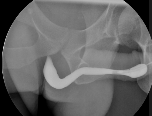
Retrograde urethrogram should be performed in male patients with pelvic trauma and blood at the urethral meatus, gross hematuria, difficulty voiding, perineal bruising, or a boggy prostate. A cystogram should be performed after ruling out urethral injury. Extravasation of contrast during these studies is suggestive of urethral or bladder injury, and Urology should be consulted [6].

Figure 2. Retrograde Urethrogram without extravasation. Case courtesy of Dr. Matt A. Morgan, Radiopaedia.org, rID: 40478.
References
[1] Manson T, O'Toole RV, Whitney A, Duggan B, Sciadini M, Nascone J. Young-Burgess classification of pelvic ring fractures: does it predict mortality, transfusion requirements, and non-orthopaedic injuries? J Orthop Trauma. 2010 Oct;24(10):603-9. PMID: 20871246.
[2] Perry K, Mabrouk A, Chauvin BJ. Pelvic Ring Injuries. [Updated 2021 Aug 6]. In: StatPearls [Internet]. Treasure Island (FL): StatPearls Publishing; 2021 Jan-. Available from: https://www.ncbi.nlm.nih.gov/books/NBK544330/
[3] Wong JM, Bucknill A. Fractures of the pelvic ring. Injury. 2017 Apr;48(4):795-802. PMID: 24360668.
[4] Griffin DR, Starr AJ, Reinert CM, Jones AL, Whitlock S. Vertically unstable pelvic fractures fixed with percutaneous iliosacral screws: does posterior injury pattern predict fixation failure? J Orthop Trauma. 2006 Jan;20(1 Suppl):S30-6; discussion S36. PMID: 16385205.
[5] Davis DD, Foris LA, Kane SM, et al. Pelvic Fracture. [Updated 2021 Aug 8]. In: StatPearls [Internet]. Treasure Island (FL): StatPearls Publishing; 2021 Jan-. Available from: https://www.ncbi.nlm.nih.gov/books/NBK430734/
[6] Uehara DT, Eisner RF. Indications for retrograde cystourethrography in trauma. Ann Emerg Med. 1986 Mar;15(3):270-2. PMID: 3511789.
[7] Ghaemmaghami V, Sperry J, Gunst M, Friese R, Starr A, Frankel H, Gentilello LM, Shafi S. Effects of early use of external pelvic compression on transfusion requirements and mortality in pelvic fractures. Am J Surg. 2007 Dec;194(6):720-3; discussion 723. doi: 10.1016/j.amjsurg.2007.08.040. PMID: 18005760.


