Stepping into Understanding: Navigating the World of Navicular Stress Fractures
Author: Zachary Brandt BS, Molly Estes MD
Orthopedic Commentary: Rodney Brandt MD
Peer-Reviewer: Brandon Godfrey MD, CAQ-SM
Final Editor: Alex Tomesch, MD, CAQ-SM
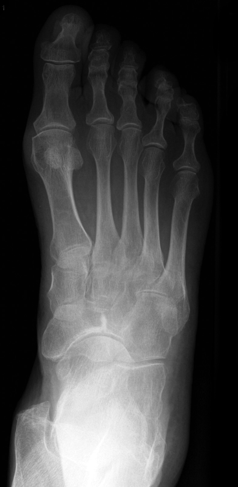
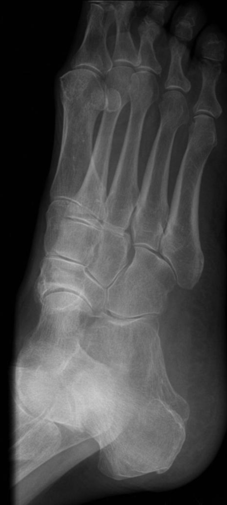
A 24-year-old male basketball player presents to the ED with right dorsal mid-foot pain after a week of daily intensive practices. The pain is worsened by use. Dorsalis pedis pulses are 2+ and the range of motion/sensation of the foot is intact.


Image 1,2. Case courtesy of Frank Gaillard, Radiopaedia.org, rID: 7202
What is the diagnosis?
Navicular stress fracture.
-
Pearl: The navicular bone is especially prone to stress fractures due to its exposure to mechanical pressure during weight-bearing activities and its relatively poor blood supply, limiting its ability to heal after stress [2,3].
What is the mechanism of injury?
Navicular stress fractures typically occur as a consequence of prolonged overuse, commonly observed after extended periods of vigorous physical activity without sufficient intervals for rest and recovery. In these situations, the navicular bone undergoes repetitive and constant strain, leading to the formation of microfractures and the creation of a vulnerable point in the foot structure [3,4].
|
Orthopedic Commentary by Rodney Brandt, MD
With a foot strike on the ground, the navicular bone experiences compression between the talus and cuneiforms. The central third of the navicular bone bears the brunt of the force during this process. This becomes significant as the central third coincides with the blood supply watershed, contributing to the occurrence of stress fractures.
|
What physical exam findings are expected?
A navicular stress fracture will typically present with milder symptoms when compared to a navicular body fracture. Symptoms include diffuse midfoot pain. Examination will reveal well-localized pain on dorsal palpation of the navicular [5].
-
Pearl: Navicular stress fractures are unique to other stress fractures in that they are more likely to present in men than women [6]. These fractures commonly exist in elite athletes such as those participating in track and field, football, and basketball.
|
Orthopedic Commentary by Rodney Brandt, MD
The nickel-sized area at the central region of the proximal dorsal navicular bone is referred to as the “N” spot. It is tender in 81 percent of patients with navicular stress fractures [7].
|
Which imaging modalities can be used?
Standard imaging protocol starts with anterior-posterior, lateral, and oblique X-rays of the foot. X-ray imaging has a low sensitivity in diagnosing this fracture, so if a navicular fracture is suspected cross-sectional imaging should be considered (but may be reserved for an outpatient setting) [3].
What is the management in the ED? When do you consult Orthopedics?
If there is suspicion of a navicular stress fracture in the ED, application of a posterior short leg splint with a stirrup or CAM boot and absolute non-weight bearing status is indicated. While in the ED it would be best to get CT imaging along with organizing close follow-up as early diagnosis leads to better outcomes.
|
Orthopedic Commentary by Rodney Brandt, MD
Early diagnosis and referral to Orthopedics, or other equivalent specialists, is key to achieving the best outcome for the patient. Operative treatment is reserved for those with six weeks of failed conservative management (eg splinting and non-weight bearing).
|
References
[1] Gaillard F, Navicular stress fracture. Case study, Radiopaedia.org (Accessed on 19 Feb 2024) https://doi.org/10.53347/rID-7202
[2] Khan KM, Brukner PD, Kearney C, Fuller PJ, Bradshaw CJ, Kiss ZS. Tarsal navicular stress fracture in athletes. Sports Med 1994: 17: 65-76.
[3] Weel, H., K. T. M. Opdam, and G. M. Kerkhoffs. "Stress fractures of the foot and ankle in athletes, an overview." Clin Res Foot Ankle 2.4 (2014): 160.
[4] Rosenbaum AJ, Uhl RL, DiPreta JA. Acute fractures of the tarsal navicular. Orthopedics. 2014 Aug;37(8):541-6. doi: 10.3928/01477447-20140728-07. PMID: 25102497.
[5] Wright AA, Taylor JB, Ford KR, Siska L, Smoliga JM. Risk factors associated with lower extremity stress fractures in runners: a systematic review with meta-analysis. Br J Sports Med. 2015 Dec;49(23):1517-23. doi: 10.1136/bjsports-2015-094828. Epub 2015 Jul 17. PMID: 26582192.
[6] Torg JS, Pavlov H, Cooley LH, Bryant MH, Arnoczky SP, Bergfeld J, Hunter LY. Stress fractures of the tarsal navicular. A retrospective review of twenty-one cases. J Bone Joint Surg Am. 1982 Jun;64(5):700-12. PMID: 7085695.
[7] Coris EE, Lombardo JA. Tarsal navicular stress fractures. Am Fam Physician. 2003 Jan 1;67(1):85-90. PMID: 12537171.


