The Gas Pedal is Connected to the Heel Bone
Author: Jasmine S. Holmes, MD (@drjazholmes)
Peer-Reviewer: Dwayne D’Souza, MD, CAQ-SM
Final Editor: Alex Tomesch, MD, CAQ-SM
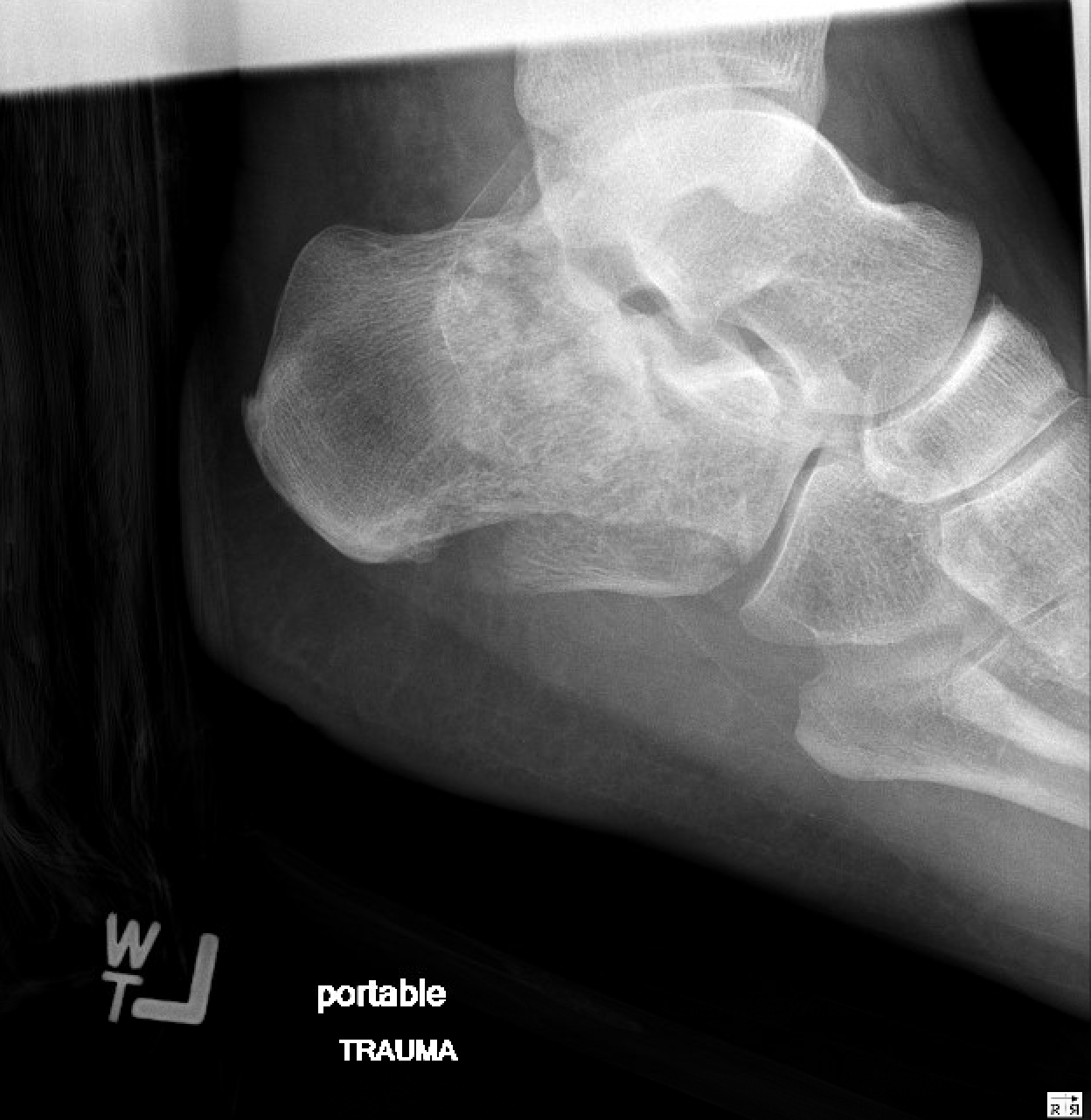
A 36-year-old male was involved in a motor vehicle collision. The patient was the restrained driver in a head-on collision at highway speeds. He experienced bilateral heel pain. Plain films of the patient's left and right calcaneus were obtained.
Image 1(a). Plain radiograph of the right calcaneus. Author's own image.
Image 1(b). Plain radiograph of the right calcaneus. Author’s own Image.

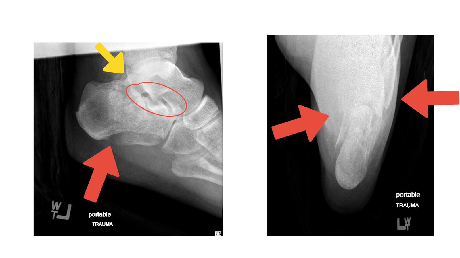
Image 1(c). Plain radiograph of left calcaneus. Author's own image.
Image 1(d). Plain radiograph of the left calcaneus. Author’s own image.
What is the diagnosis?
Right comminuted calcaneal fracture Image 1(a) and 1(b) with annotations to show the findings (red arrows).

Left comminuted and displaced fractures of the calcaneus (red arrows) and talus (yellow arrow) with at least subluxation at the subtalar joints (red circle), Image 1(c) and 1(d).

Image 2. Image courtesy of EBM Consult, LLC
What is the mechanism of injury?
High energy impact of the heel on a hard surface. Examples include a fall from a 2nd story building or a high-energy motor vehicle collision.
What physical exam findings are expected?
On examination, calcaneus fractures will present with pain, edema, and ecchymoses at the site. The patient is unable to bear weight. Suspicion should be raised significantly if there is plantar ecchymosis through the plantar arch of the foot [2]. Look for skin tenting, blistering, and skin breakdown at the heel, for this may be an indication of a tongue-type fracture.
Which imaging modalities can be used?
Plain radiographs of the heel, lateral, and axial views.
Computed tomography (CT) scan to provide better visualization of subtalar joint and assess fracture pattern and injuries [4]. It can accurately evaluate fracture lines, dislocation, and comminution, utilizing multiplanar reformation [5]. CT also gives improved visualizations of the surrounding tissues and can show tendon entrapment or dislocation [3].
What is the management in the ED?
Immobilization with a splint, usually with a bulky dressing. The patient should be non-weight-bearing.
When do you consult Orthopedics?
Emergent Orthopedics consultation is needed for intra-articular and displaced fractures. Tongue-type fractures must be seen in HOURS of initial injury due to complications from skin tenting [6].
Outpatient Orthopedic follow-up (after compressive dressing and casting) for extra-articular fractures [6].
References
[1] Isaacs JD, Baba M, Huang P, et al. The Diagnostic Accuracy of Böhler’s Angle in Fractures of the Calcaneus. The Journal of Emergency Medicine. 2013;45(6):879-884.
[2] Davis D, Seaman TJ, Newton EJ. Calcaneus Fractures. [Updated 2023 Jul 31]. In: StatPearls [Internet]. Treasure Island (FL): StatPearls Publishing; 2023 Jan-. Available from: https://www.ncbi.nlm.nih.gov/books/NBK430861/
[3] White EA, Skalski M, Matcuk GR, et al. Intra-articular tongue-type fractures of the calcaneus: anatomy, injury patterns, and an approach to management. Emergency Radiology. 2018;26(1):67-74.
[4] Johnson PT, Fayad LM, Fishman EK. Sixteen-slice CT with volumetric analysis of foot fractures. Emergency Radiology. 2006;12(4):171-176.
[5] Gilmer PW, Herzenberg JE, J. Lawrence Frank, Silverman PM, Martinez S, J. Leonard Goldner. Computerized Tomographic Analysis of Acute Calcaneal Fractures. Foot & ankle. 1986;6(4):184-193.
[6] Abramson Tiffany. Calcaneus Fracture. In: Mattu A and Swadron S, ed. CorePendium. Burbank, CA: CorePendium, LLC. https://www.emrap.org/corependium/chapter/receLoADvxROW3b2u/Calcaneus-Fracture#h.1hmsyys. Updated May 10, 2022. Accessed November 30, 2023.


