Twisting Tales: Unraveling the Complexities of the Galeazzi Fracture
Author: Zachary Brandt BS, Molly Estes MD
Orthopedic Commentary: Rodney Brandt MD
Peer-Reviewer: Mark Hopkins, MD, CAQ-SM
Final Editor: Alex Tomesch, MD, CAQ-SM
A 30-year-old male presents to the ED after a motorcycle accident. You find a wrist deformity on his exam. There is a 2+ radial pulse and motor/sensation of the hand is intact.


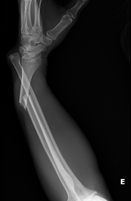
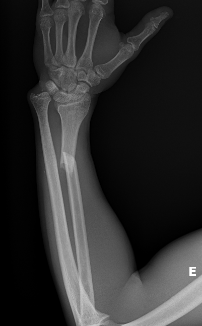
Image 1. Plain radiograph of the right forearm. Case courtesy of Leonardo Lustosa, Radiopaedia.org, rID: 166236
What is the diagnosis?
Distal radius fracture with a dislocation of the distal radioulnar joint (DRUJ), which describes a Galeazzi fracture-dislocation.
-
Pearl: In most cases, the DRUJ injury is not as clearly demonstrated, so additional findings to look for are ulnar styloid base fracture, dislocation/subluxation of the radius relative to ulna on the lateral view, and shortening of the radius by more than 5mm [1].
|
Orthopedic Commentary by Rodney Brandt, MD
There is a need (on these images) for a true lateral and anteroposterior x-ray of the wrist, NOT an oblique. Although Galeazzi fractures are uncommon in the pediatric population, one study observed that 41% of Galeazzi fractures went unrecognized [2]. A true AP and lateral x-ray help reduce this common error.
An increased index of suspicion should exist when a radial shaft fracture is within 7.5cm of the distal radius. This has a 55% association with DRUJ injury [3].
|
What is the mechanism of injury?
Galeazzi fractures occur due to an axial load onto an outstretched hand. If the arm is supinated on impact, an apex volar radial shaft fracture is expected to have an associated volar dislocated ulna within the DRUJ. In contrast, if the arm is pronated on impact, an apex dorsal angulated radial shaft fracture is expected along with a dorsal dislocation of the ulna within DRUJ [4]. See images here.
What physical exam findings are expected?
On physical exam, there will be swelling of the hand and wrist along with pain upon flexion and extension of the wrist [5]. As always, but especially given the extreme angulation of these fractures, be sure to check neurovascular status.
-
Pearl: These can be associated with an anterior interosseous nerve (AIN) palsy [6]. The AIN is the pure motor branch of the median nerve and innervates the flexor pollicis longus and flexor digitorum profundus to the second digit; thus, palsy of AIN can lead to weak opposition between the first and second digit.
|
Orthopedic Commentary by Rodney Brandt, MD
Gentle palpation of the distal ulna will elicit pain due to ligamentous injury of the DRUJ.
|
Which imaging modalities can be used?
Standard anteroposterior and true lateral radiograph films should be obtained of both the elbow and the wrist [7]. To assess for DRUJ disruption either contralateral films can be obtained or a bilateral axial computed tomography (CT).
What is the management in the ED? When do you consult Orthopedics
Adolescent patients have been shown to have successful outcomes with closed reduction and further assessment of the DRUJ under fluoroscopy. For adult patients, however, open reduction and internal fixation (ORIF) is necessary [7]. Splinting should be performed in the ED for all patients, with orthopedics consulted for close follow-up (generally outpatient, depending on institutional protocols). As always, if there are signs of neurovascular compromise or there is concern for compartment syndrome, an emergent consult would be warranted.
|
Orthopedic Commentary by Rodney Brandt, MD
For pediatric patients, the most important part of the reduction of Galeazzi fractures is the reversal of the mechanism of injury.
In adult patients, splinting should be performed in situ and follow-up with orthopedics urgently as they require operative treatment [7]. Non-operative treatment has resulted in unsatisfactory outcomes in 92% of patients [3].
|
References
[1] Lustosa L, Galeazzi fracture-dislocation. Case study, Radiopaedia.org (Accessed on 25 Nov 2023) https://doi.org/10.53347/rID-166236
[2] Walsh HP, McLaren CA, Owen R. Galeazzi fractures in children. J Bone Joint Surg Br. 1987 Nov;69(5):730-3. doi: 10.1302/0301-620X.69B5.3680332. PMID: 3680332.
[3] David E. Ruchelsman, Keith B. Raskin, Michael E. Rettig, CHAPTER 22 - Galeazzi Fracture-Dislocations, Editor(s): David J. Slutsky, A. Lee Osterman, Fractures and Injuries of the Distal Radius and Carpus, W.B. Saunders, 2009, Pages 231-239, ISBN 9781416040835, https://doi.org/10.1016/B978-1-4160-4083-5.00024-X.
[4] Alajmi T. Galeazzi Fracture Dislocations: An Illustrated Review. Cureus. 2020 Jul 24;12(7):e9367. doi: 10.7759/cureus.9367. PMID: 32850236; PMCID: PMC7444983.
[5] Andrew D. Perron, Robert E. Hersh, William J. Brady, Theodore E. Keats, Orthopedic pitfalls in the ED: Galeazzi and Monteggia fracture-dislocation, The American Journal of Emergency Medicine, Volume 19, Issue 3, 2001, Pages 225-228, ISSN 0735-6757,https://doi.org/10.1053/ajem.2001.22656.
[6] Galanopoulos I, Fogg Q, Ashwood N, Fu K. A widely displaced Galeazzi-equivalent lesion with median nerve compromise. BMJ Case Rep. 2012 Aug 18. 2012
[7] Atesok, Kivanc I. MD; Jupiter, Jesse B. MD; Weiss, Arnold-Peter C. MD. galeazzi Fracture. American Academy of Orthopaedic Surgeon 19(10):p 623-633, October 2011.
[8] Tsismenakis T, Tornetta P 3rd. Galeazzi fractures: Is DRUJ instability predicted by current guidelines? Injury. 2016 Jul;47(7):1472-7. doi: 10.1016/j.injury.2016.04.003. Epub 2016 Apr 20. PMID: 27138839.


