
- Details
- Hits: 1069

Regional Anesthesia Upper Extremity
December 2024
Final Reviewer: Justine Ko, MD, CAQ-SM
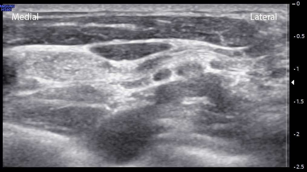
Image 1: Author’s own image.

Image 2: Demonstration of the ultrasound technique for this case. Author’s own image.
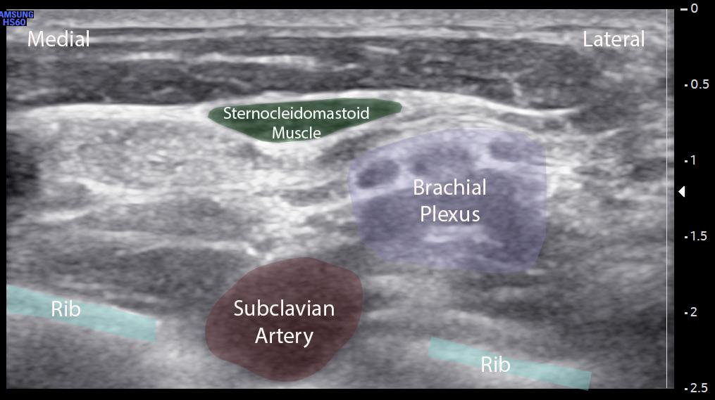
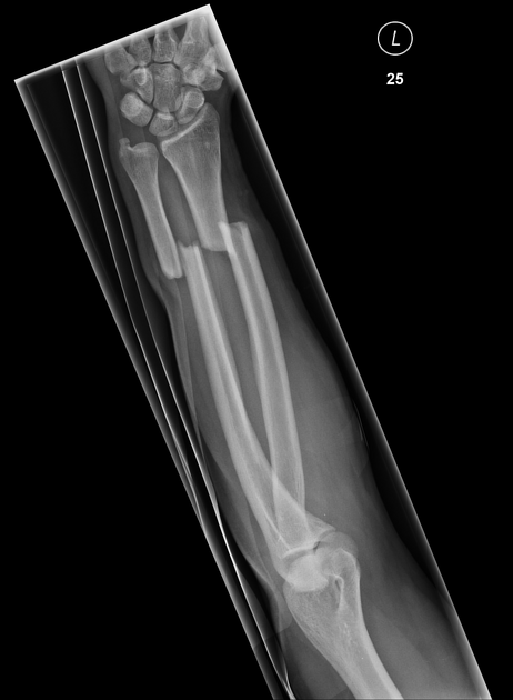
Image 4: Both bone forearm fracture with displacement, Image courtesy of Jones J, Niknejad M, Bickle I, et al., Radiopaedia.org, rID: 43378
What is the patient positioning and positioning of the ultrasound for this procedure?
The supraclavicular block can be performed with the patient either sitting up, partially reclined, or with the patient supine. The author prefers to perform with the patient sitting up, with the operator approaching from a posterolateral position. The ultrasound should be placed across from the operator’s field of view.
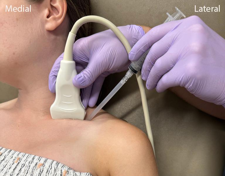
Image 2. Positioning to perform supraclavicular nerve block, author’s image
How can you identify the desired location for the supraclavicular nerve block. What are key structures to identify?
We recommend starting by placing the transducer in a roughly transverse position just above the clavicle. Angle the transducer upright. The supraclavicular nerve block is essentially a brachial plexus nerve block. Identify the subclavian artery (and if possible, subclavian vein). The brachial plexus appears as a collection of hypoechoic “grape-like’ structures, typically lateral and superficial to the subclavian artery. Apply color doppler to the area to confirm the hypoechoic structures are the brachial plexus, and not additional blood vessels. Also identify lung slide/pleura, and note the relative depth of these structures.
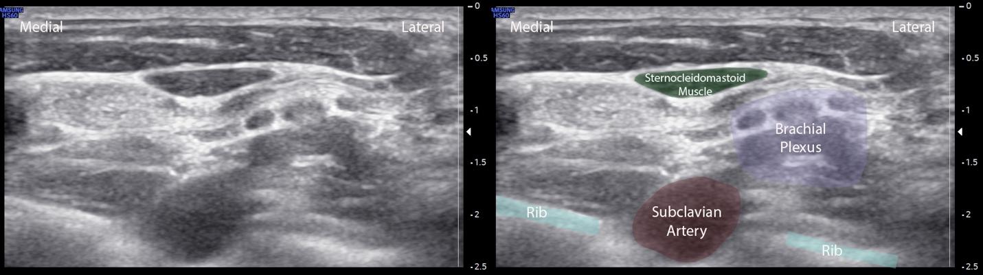
Image 5. Ultrasound image of the supraclavicular space including the subclavian artery and brachial plexus, author’s image
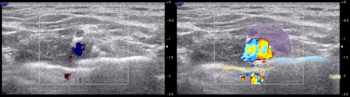
Image 6. Ultrasound image of color doppler confirming subclavian artery vs brachial plexus, author’s image
What is the distribution of anesthesia for a supraclavicular nerve block?
The supraclavicular nerve block is a brachial plexus block that results in reliable anesthesia of most of the upper extremity. The supraclavicular approach is typically performed at the level of the trunks of the brachial plexus. This results in anesthesia of the median, ulnar, and radial peripheral nerves distally and can be used for injuries from the mid-humerus to distal. It does not reliably anesthetize the intercostobrachial nerve (medial upper arm), nor does it reliably affect the suprascapular nerve, so it should not be used for purely shoulder or glenohumeral pathology.
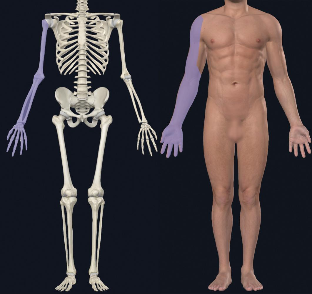
Image 7. Regional anesthesia distribution of the supraclavicular nerve block, author’s image.
What is the target for your needle for the block?"
Insert your needle from lateral to medial. Advance the needle through the soft tissue and deposit the anesthetic adjacent to the brachial plexus. Unlike other nerve blocks, do not attempt to create a “halo” around the brachial plexus due to the risk of injuring nearby structures, including the subclavian artery and lung pleura. For this block, the goal is to deposit the anesthetic near the brachial plexus and allow it to diffuse. Do not inject directly into the nerve bundles. If you feel resistance with the syringe, this suggests that the needle is in the nerve bundle or another important structure.
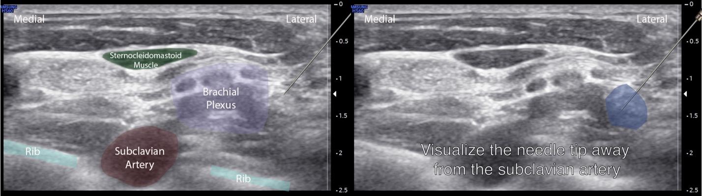
Image 8. Supraclavicular nerve injection, author’s own image
What is the patient positioning and positioning of the ultrasound for this procedure?
The supraclavicular block can be performed with the patient either sitting up, partially reclined, or with the patient supine. The author prefers to perform with the patient sitting up, with the operator approaching from a posterolateral position. The ultrasound should be placed across from the operator’s field of view.
Image 2. Positioning to perform supraclavicular nerve block, author’s image
How can you identify the desired location for the supraclavicular nerve block. What are key structures to identify?
We recommend starting by placing the transducer in a roughly transverse position just above the clavicle. Angle the transducer upright. The supraclavicular nerve block is essentially a brachial plexus nerve block. Identify the subclavian artery (and if possible, subclavian vein). The brachial plexus appears as a collection of hypoechoic “grape-like’ structures, typically lateral and superficial to the subclavian artery. Apply color doppler to the area to confirm the hypoechoic structures are the brachial plexus, and not additional blood vessels. Also identify lung slide/pleura, and note the relative depth of these structures.
Image 5. Ultrasound image of the supraclavicular space including the subclavian artery and brachial plexus, author’s image
Image 6. Ultrasound image of color doppler confirming subclavian artery vs brachial plexus, author’s image
What is the distribution of anesthesia for a supraclavicular nerve block?
The supraclavicular nerve block is a brachial plexus block that results in reliable anesthesia of most of the upper extremity. The supraclavicular approach is typically performed at the level of the trunks of the brachial plexus. This results in anesthesia of the median, ulnar, and radial peripheral nerves distally and can be used for injuries from the mid-humerus to distal. It does not reliably anesthetize the intercostobrachial nerve (medial upper arm), nor does it reliably affect the suprascapular nerve, so it should not be used for purely shoulder or glenohumeral pathology.
Image 7. Regional anesthesia distribution of the supraclavicular nerve block, author’s image.
What is the target for your needle for the block?
Insert your needle from lateral to medial. Advance the needle through the soft tissue and deposit the anesthetic adjacent to the brachial plexus. Unlike other nerve blocks, do not attempt to create a “halo” around the brachial plexus due to the risk of injuring nearby structures, including the subclavian artery and lung pleura. For this block, the goal is to deposit the anesthetic near the brachial plexus and allow it to diffuse. Do not inject directly into the nerve bundles. If you feel resistance with the syringe, this suggests that the needle is in the nerve bundle or another important structure.
Image 8. Supraclavicular nerve injection, author’s own image